Making up your mind
Inside UC Berkeley’s powerful new brain scanner. Published in California Monthly magazine.
On the northwest side of campus, in the middle of Wellman Court, sits a small trailer painted cornflower blue, surrounded by a little yard—a patch of grass, in truth—and a few meters of high fencing. Its diminutive size and modern angular look give it the appearance of a comically oversized architect’s model.
But appearances can be deceptive. This modest structure houses a piece of cutting-edge technology—a $5-million magnetic resonance imaging (MRI) scanner that can actually “watch your brain think.” Built last November, the Henry H. Wheeler Jr. Brain Imaging Center, as the blue trailer prefers to be called, will provide detailed snapshots of mental activity that will help neuroscientists “figure out how the brain is related to the mind,” in the words of psychology professor Richard Ivry.
This state-of-the-art machine places Berkeley neuroscientists in a better position than most to answer that question because it is one of the only MRI scanners in the world devoted solely to brain research. Most MRI brain imaging studies rely on hospital scanners, which means that researchers like Ivry have to wait until after hours before they can get to them. By the time they have set up their experiments and are ready to begin scanning, it might be as late as 10 p.m., according to Corey Goodman, head of Berkeley’s Helen Wills Neuroscience Institute, a grouping of around 40 faculty members from various departments, all with an interest in the brain. “So all these studies that you see in magazines and newspapers of the normal human brain are actually about the very sleepy human brain,” jokes Goodman, who says that the Brain Imaging Center is the first of three “technology cores” planned for the Neuroscience Institute (the other two being a Neurogenomics Center and a Molecular Imaging Center).
There is a palpable excitement among Berkeley neuroscientists about their new machine, and not just because it will allow them to keep more sociable hours. Powered by a huge, liquid-helium, superconducting magnet, the custom-built scanner has a magnetic field strength of 4 Tesla—nearly three times the strength of a standard hospital MRI, and 10,000 times the strength of the Earth’s own magnetic field. It is not only more powerful than a typical MRI, it’s also much more flexible, so it can be customized for just about any experiment the researchers dream up. “We wanted a machine that a medical technician would never come near, one that’s highly programmable, that requires physicists and computer scientists to be on hand,” says Goodman. “What have we got at Berkeley? We may not have hospital beds, but we have physics, chemistry, computer science, math, statistics, engineering—we have incredible physical and quantitative sciences around us, and these people are very interested in helping us develop new-generation methods.”
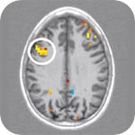
The current method of choice in brain imaging is functional MRI (fMRI). A standard MRI scan uses its strong magnetic field to line up the atoms of water molecules in the brain, which are then detected by using radio frequency pulses. A sharp, three-dimensional image of the brain is then constructed from these signals. Functional MRI goes one step further, by also tracking blood flow in the brain. As neurons become active, they need additional oxygen-rich blood, and an MRI scanner is sensitive enough to detect the difference between hemoglobin molecules carrying oxygen and those without, allowing it to monitor brain activity—albeit indirectly.
With its incredibly powerful field strength, the new scanner at Berkeley can pinpoint brain activity right down to the nearest millimeter of its happening and within a second of its occurring—the highest resolution of any machine so far approved for use on humans. “And we haven’t reached the limit of spatial or temporal resolution,” says a confident Mark D’Esposito, director of the Brain Imaging Center.
D’Esposito and his colleagues are particularly interested in using the scanner to understand the “executive” functions of the brain—higher-level abilities such as handling several tasks at once, responding to new information, and making decisions about how to act. Executive functions are closely associated with the prefrontal cortex—the most highly developed area of the human brain, taking up one-third of the cortex, the outer layer of the brain. (The prefrontal area takes up just one-tenth of a chimp’s cortex.) “It’s the most complicated area to study—how we can voluntarily decide to do one thing or another,” says D’Esposito, undaunted by the challenge. “The back of the brain without the frontal lobes would just be out of control, reacting to everything reflexively. The frontal lobes only act on information that’s relevant, that guides your actions,” he explains.
D’Esposito is one of two clinical neurologists in Berkeley’s psychology department—the other is Bob Knight—and when they aren’t busy with their research, they continue to see patients at the neurology clinic of the Veterans Administration Hospital in Martinez. Both appointments are highly unusual for a psychology department, but reflect Berkeley’s focus on the brain processes that underlie our thoughts, feelings, and actions. Most of Knight’s patients have damage to their prefrontal cortex, and he studies them in part to understand how the brain works. He describes a typical patient: “They’re distractible; they tend to drift off set. They have trouble with planning, and they tend to get stuck in the present. In routinized testing they score normal, but if you give them tests that require thinking—any creativity or flexibility—they bomb.”
When the prefrontal cortex is working properly, it uses information coming in from the senses in a “smart” way, explains Knight. By paying attention to those things we consider important, and ignoring those we don’t, we can manage our mental resources more efficiently. Without the help of the prefrontal cortex, all information from the senses assumes equal importance, and the result is confusion. People with schizophrenia, who share many of the symptoms of Knight’s patients, often have decreased activity in this part of the brain, and he hopes that his research might eventually result in better treatments for this and other psychiatric diseases that involve the prefrontal cortex, including attention-deficit disorder.
“The prefrontal cortex allows you to amp up attention to what you’re interested in, and to inhibit what you’re not interested in,” continues Knight, who has spent years monitoring this “amping up” and “damping down,” using electrodes attached to the scalp to record electroencephalograph (EEG) signals, the electrical impulses that accompany brain activity. He especially wants to examine how it is that healthy people are able to rapidly shift their focus and respond to something unexpected from outside their field of attention. An example of this is the “cocktail party effect,” a phenomenon psychologists have known about for years. At a noisy party, you effortlessly zero in on one conversation, filtering out the background noise. But should you suddenly hear your own name in a different conversation, you immediately become aware of it and turn around. While you were listening to one conversation, unbeknownst to you, part of your brain was subtly eavesdropping on the rest of the party.
Knight is trying to repeat his EEG recordings in the new MRI scanner—a feat that has never before been accomplished, and which he has spent all summer trying to pull off. By combining the two methods, EEG and fMRI, he’s hoping to get the best of both worlds—the incredibly sharp resolution of the MRI scanner, together with the speed of the EEG, to produce the most precise information yet on the time and location of brain activity. It hasn’t been easy. Measuring tiny electric currents in a huge magnetic field is tricky—the slightest twitch of the head, even the tiny movement caused each time a subject’s heart beats, induces a small electric current in the EEG wires. Knight has been experimenting with carbon wires and electrodes to reduce distortions in the magnetic field and, despite the technical hurdles, is confident that he will be ready to begin the simultaneous recordings in the fall. “It’s definitely worth the effort we’re putting into it,” he believes.
One of Knight’s first experiments using the scanner will involve two different sets of patients with frontal lobe damage—one with damage to their lateral prefrontal cortex, and the other with damage to their orbital prefrontal cortex, the part of the brain that sits just behind the eyes. The second set of patients has a quite different set of symptoms from the first—their intellect is entirely unaffected, but their social skills are severely limited. The most celebrated example of such a case is that of Phineas Gage, a Vermont railroad foreman, injured in 1848 by an explosion that blasted an inch-thick iron rod through his left eye and out the top of his skull. Despite a remarkable physical recovery, this amiable, hard-working man was turned, in an instant, into a foul-mouthed, impulsive, unreliable rogue. “Gage,” his friends said, “was no longer Gage.” Ladies were advised to stay well away.
When Gage’s doctor first reported the case, few physicians believed him, but today those symptoms are recognized as typical of orbital prefrontal damage. “Such patients don’t get good ‘reads’ on people, and they get into all kinds of social mischief because they make bad decisions. They have a horrible time interacting in the social world, and they get ostracized because of their impulsive and inappropriate behavior,” says Knight. He predicts that his two sets of patients will react quite differently in his experiment, which will measure their responses to novelty while their attention is focused elsewhere. “If I were to make a loud and unexpected clap during an experiment, someone with lateral frontal damage would show a lack of behavioral and physiological response. If I did the same thing with someone with orbital frontal damage, they’d show heightened activity in the auditory part of their brain; they’d over-respond,” he says.
Victorian doctors may have been incredulous that a single part of the brain could control social behavior. But around the time of Gage’s accident, there were those who argued that all human traits could be traced to the pattern of lumps and bumps on the skull. Phrenology, as that school of thought was known, has long since been abandoned, but some now charge that cognitive neuroscientists like Knight, D’Esposito, and Ivry are essentially modern-day phrenologists, tracing human behaviors to this or that part of the brain. Walter Freeman, a professor of the graduate school whose office is just yards away from the Brain Imaging Center, is one such critic. He points out that all areas of the brain are active to some degree all of the time. So with fMRI, control images of the brain “doing nothing” must be subtracted to see which parts of the brain are most active during a task; this approach, he argues, inevitably leads to an artificially segmented view of the brain. Freeman believes that only a whole-brain approach—one that looks at the entire pattern of activity in the brain—will ultimately tell us how brains work.
D’Esposito is quick to defend fMRI from those who would compare it to phrenology. “A lot of the work now done with imaging is starting to move away from [a localized approach]. It’s moving more towards how things work rather than where in the brain things work.” It’s all about designing the right sort of experiments, says D’Esposito. “Imaging got really clever when cognitive psychologists started to bring in their sophisticated methods.” Nowadays, cognitive neuroscientists don’t just scan the brain, he says, they devise subtle experiments to compare different psychological theories of how the mind works.
Rich Ivry gives an example: “The most basic memory question that psychologists have been asking forever and ever is: Do we not remember something because we can’t retrieve it, or because we never encoded it properly? They’re two fundamentally different psychological models.” In an experiment to decide between them, people were scanned while they were shown a list of things to remember. When they came out of the scanner they were given a memory test. “There was indeed a difference between [the scans for] items that were later remembered and items that were forgotten, which means that at least some of the problem [of forgetting] has to do with an encoding deficit.”
Yet Ivry acknowledges that even the smartest experiments, using the most precise brain-imaging techniques, are prone to over-interpretation. “The problem with most imaging studies is that you have a correlation between this task and this part of the brain being active, but it’s very hard to say that that activity is essential for doing that task,” he says.
There are others who say that the limitations of brain imaging are much more serious than neuroscientists realize. Some philosophers question the very idea that science, which describes the physical world, could ever describe such subjective experiences as “seeing red” or “tasting chocolate,” which are part of the mental world. This “mind-body” problem of philosophy was first expressed by 17th-century philosopher René Descartes, who would have said that a map of the brain could tell us no more about the mind than a map of the Earth could tell us about Heaven and Hell. Four centuries later, most of us still have a hard time comprehending how a large piece of gray, fatty tissue can produce something as intangible as the mind.
But not all philosophers worry about it. World-renowned Berkeley professor John Searle dismisses the conundrum as erroneous and confused. “I think the mind-body problem is solved by saying that, as a matter of fact, brains do it—they cause consciousness,” says Searle, who adds that “consciousness is as much a biological phenomenon as digestion or photosynthesis.” He does admit, however, that conscious states are certainly unusual in that they “only exist insofar as they’re experienced by some human or animal subject.”
Such concerns could not be further from the thoughts of the many researchers on campus who have already contacted D’Esposito to express their enthusiasm about the new scanner and the possibilities it presents. So far, interest has come from vision sciences, social psychology, and education, and D’Esposito is thrilled at the prospect of collaborating with people from numerous disciplines. “It’s early yet, and we haven’t even put the word out in a big way. When it really gets out there, and the physical sciences start participating, there are going to be questions that I haven’t even imagined,” he predicts.

